Disease progression in most pathological conditions, such as atherosclerosis, cancer, orthopedic disorders and neurodegenerative diseases is accompanied with alterations in the mechanical properties of affected tissues. For instance, the leading cause of death, myocardial infarction (MI) is caused by rupture of mechanically compromised vulnerable atherosclerotic plaques. In many neurodegenerative disorders, such as Alzheimers disease, changes in the biomechanical properties of amyloid fibrils are believed to be responsible for the proliferation, growth, and propagation of amyloid plaques. At the cellular level, altered mechanical properties of the tumor microenvironment have been shown to regulate malignant transformation and cancer cell proliferation independent of biochemical cues. Therefore, the significant evidence on the role of mechanical factors on disease initiation and progression calls for development of novel technologies for biomechanical evaluation of tissue in situ. Traditionally, a number of micro-indentation devices and scanning probe microscopes have been commercialized to meet the need for micromechanical testing. These instruments however probe superficial depths (~10-20mm) over a small field of view (~100mm), require several hours of measurement time, are costly (>$250,000) and are unsuitable for tissue assessment in situ.
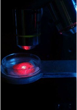
Our group has developed a new optical approach, Laser Speckle Rheology (LSR), that measures the biomechanical properties of tissues, biofluids and biomimetic scaffolds with microscale resolution. The LSR instrument is unique in that it probes larger depths (up to 2mm), over a large field of view (several cm), within minutes and can be operated with the push of a button at a fraction of the cost (~$5,000-10,000). Moreover, since LSR does not contact or manipulate the sample during measurement, it offers several distinct advantages for operation in sterile environments where sample contamination by micro-indentation devices may pose a significant problem. In LSR, an inexpensive laser source (similar to a laser pointer) illuminates the sample and laser speckle scattered from the sample is measured using a CMOS camera. By analyzing intensity fluctuations of the laser speckle over time, the biomechanical properties of the sample can be quantified. LSR is performed using a modified microscope instrument or can be conducted via miniature endoscopes or needles to assess deep tissues and internal organs in a minimally invasive manner, opening several opportunities for research and clinical investigations.
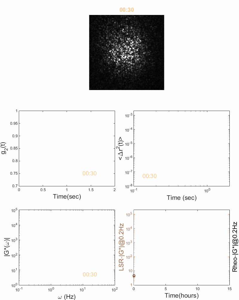
Laser Speckle Rheology quantifies changes in viscoelastic properties of materials. For instance, in a PDMS gels, speckle fluctuations slow down due to polymer molecules cross-linking during thermal curing. By Analyzing the time-lapse speckle fluctuations, LSR provides the trace of viscoelastic modulus, G*(ω), as a function of curing time, which matches the trace measured by a conventional rheometer.
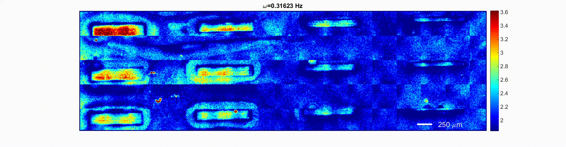 Mapping the frequency-dependent viscoelastic modulus of a micro-fabricated PDMS-PEG gel phantom. Each frame of this media file shows the spatially-resolved modulus, G, at several consecutive oscillation frequencies, ranging from ω=0.3-100 Hz. The bars in successive columns are 1 mm long and 200, 150, 100, and 80 µm wide, respectively. The color-bar scale adjusted at each frame to provide the highest visual contrast between the soft PEGDA background and the stiff PDMS bars. The 80 µm wide PDMS bars evolved from barely visible at low frequencies to fully resolved and highly contrasted from the soft PEGDA background at higher frequencies within the G* color maps. The moduli range from 10 kPa – 1 MPa, and are represented by blue to red hues. Significant contrast is observed between stiff PDMS bars and the background PEG gel at all length-scales.
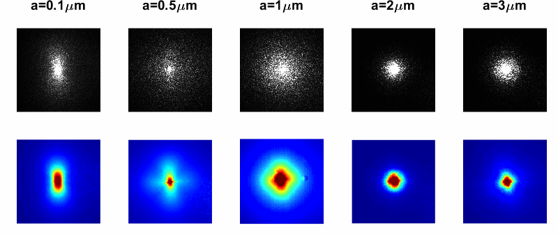
Particle sizing from diffuse scattered light. The figure shows laser speckle patterns of mono-disperse polystyrene microspheres of assorted sizes, collected through a parallel linear polarizer. Time averaging of speckle patterns yields the two-dimensional diffuse reflectance of the material. The diffuse reflectance at certain polarization angles exhibits an azimuth angle dependence, that depends on the average scattering size, a. For instance, as particle size increases, the diffuse reflectance evolves from a bi-lobular pattern to a clover-like shape.
Relevant Publications
Laser Speckle Rheology for evaluating the viscoelastic properties of hydrogel scaffolds. Hajjarian Z, Nia HT, Ahn S, Grodzinsky AJ, Jain RK, Nadkarni SK. Sci Rep. 2016 Dec 1;6:37949. doi: 10.1038/srep37949. PMID: 27905494
Estimation of particle size variations for laser speckle rheology of materials. Hajjarian Z, Nadkarni SK. Opt Lett. 2015 Mar 1;40(5):764-7. doi: 10.1364/OL.40.000764. PMID: 25723427
Correction of optical absorption and scattering variations in Laser Speckle Rheology measurements. Hajjarian Z, Nadkarni SK. Opt Express. 2014 Mar 24;22(6):6349-61. doi: 10.1364/OE.22.006349. PMID: 24663983
Evaluation and correction for optical scattering variations in laser speckle rheology of biological fluids. Hajjarian Z, Nadkarni SK. PLoS One. 2013 May 21;8(5):e65014. PMID: 23705028
Evaluating the viscoelastic properties of tissue from laser speckle fluctuations. Hajjarian Z, Nadkarni SK. Sci Rep. 2012;2:316. doi: 10.1038/srep00316. Epub 2012 Mar 16. PMID: 22428085