laser speckle rheological microscopy (SHEAR)

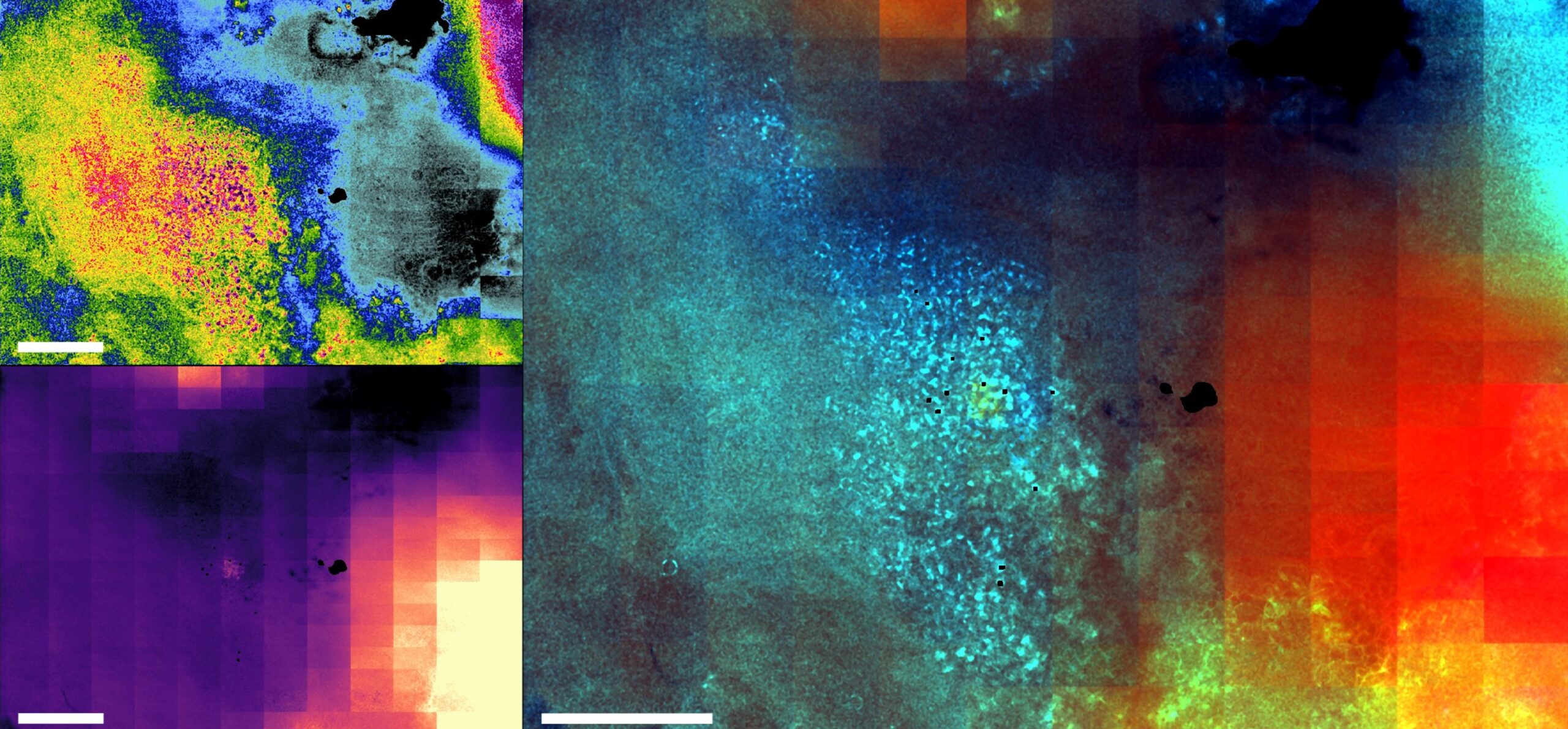
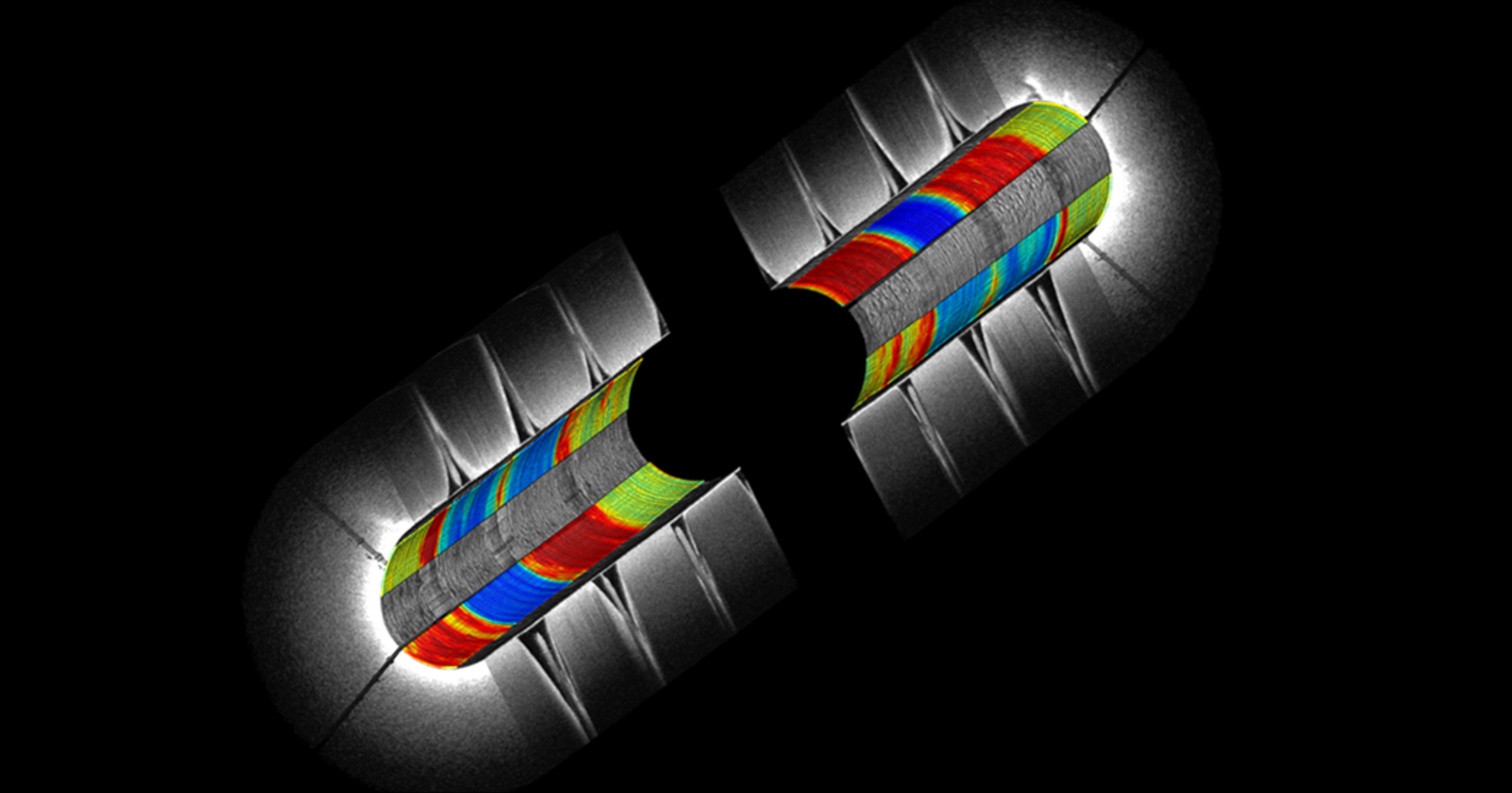
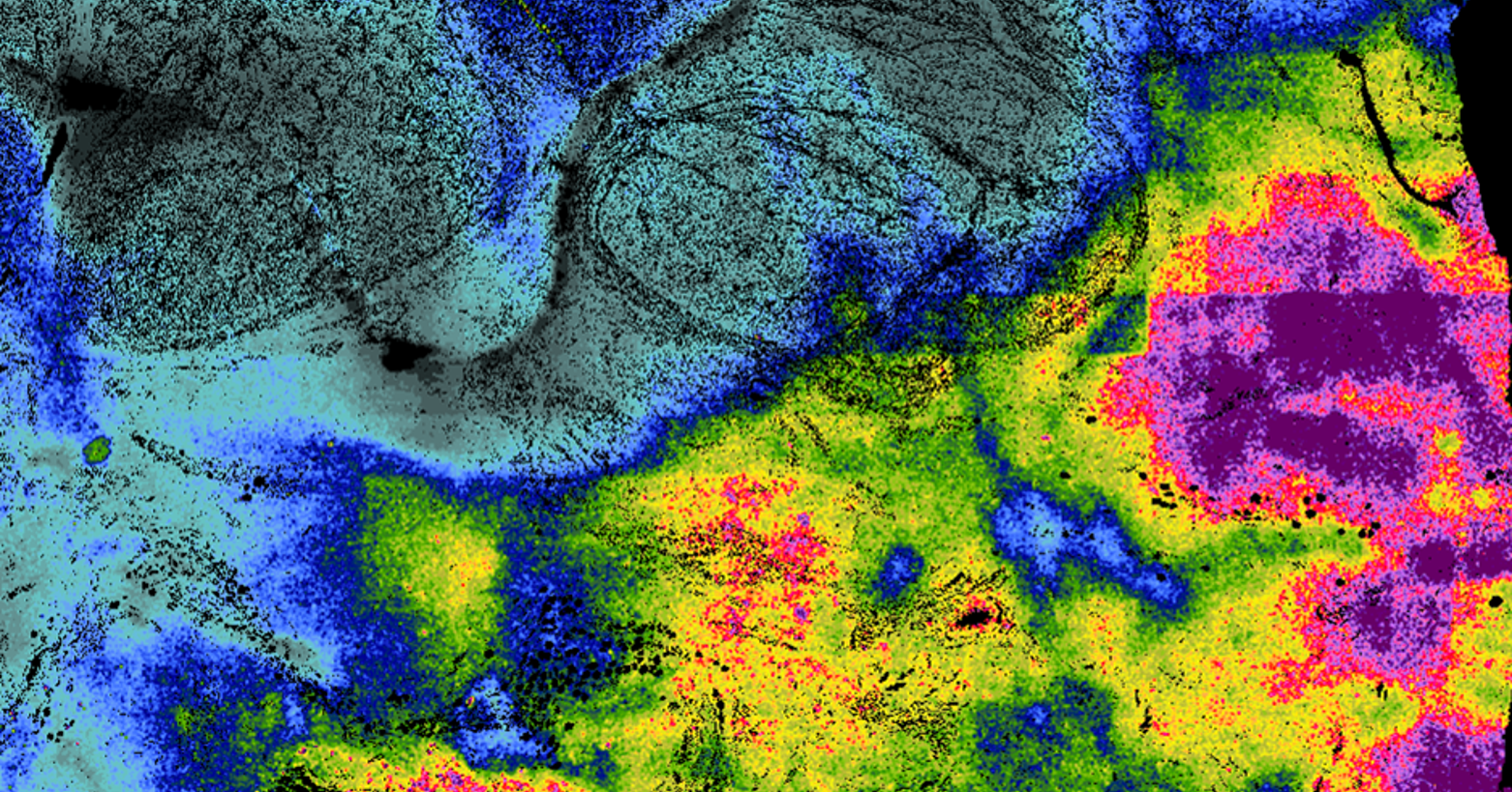
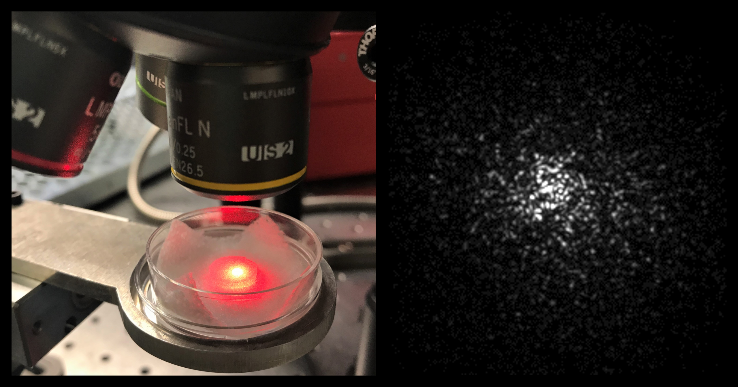
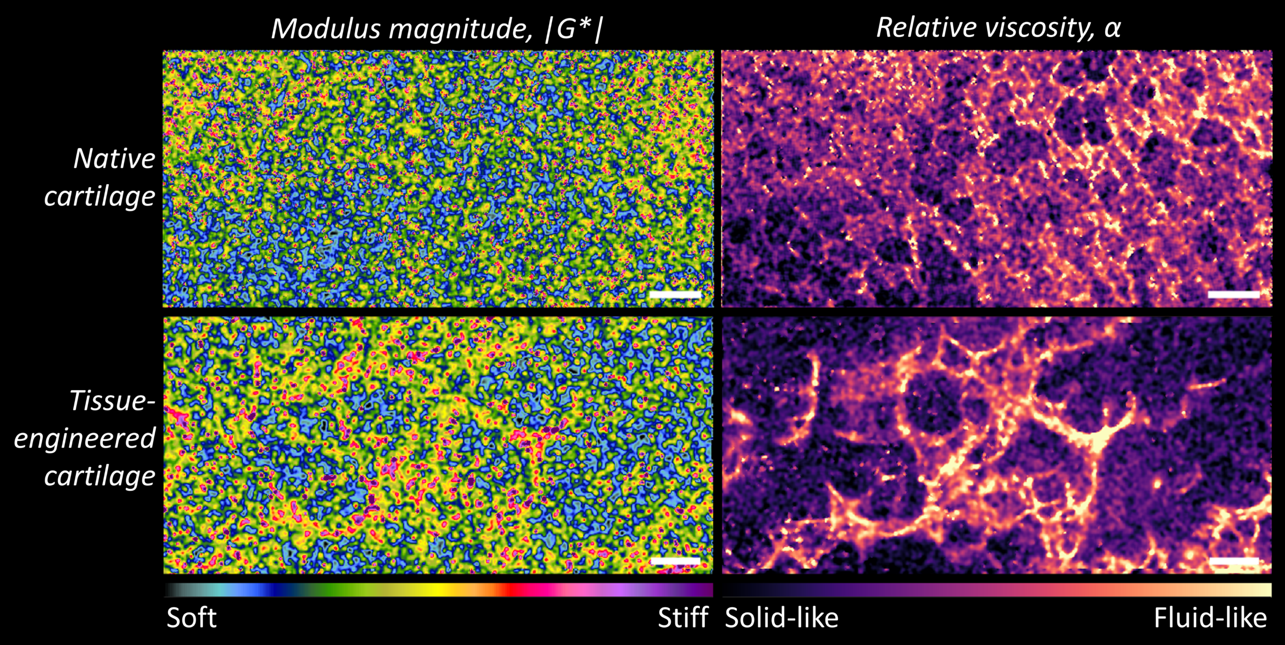
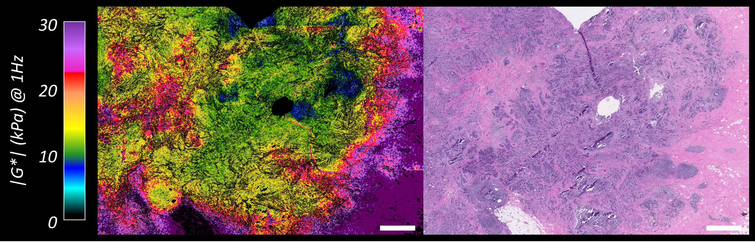
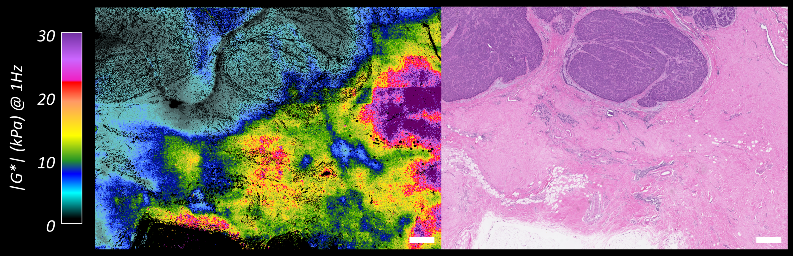
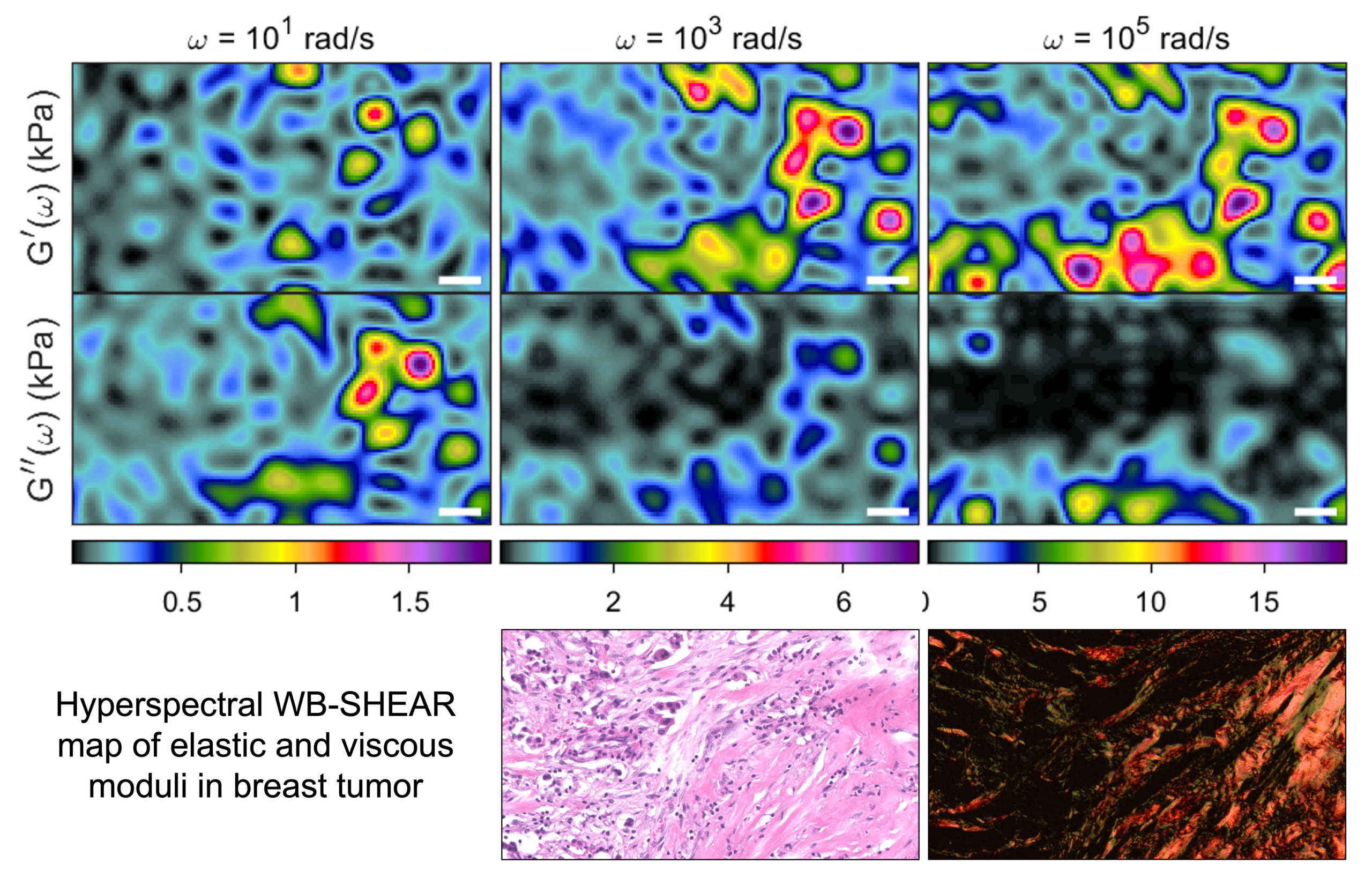
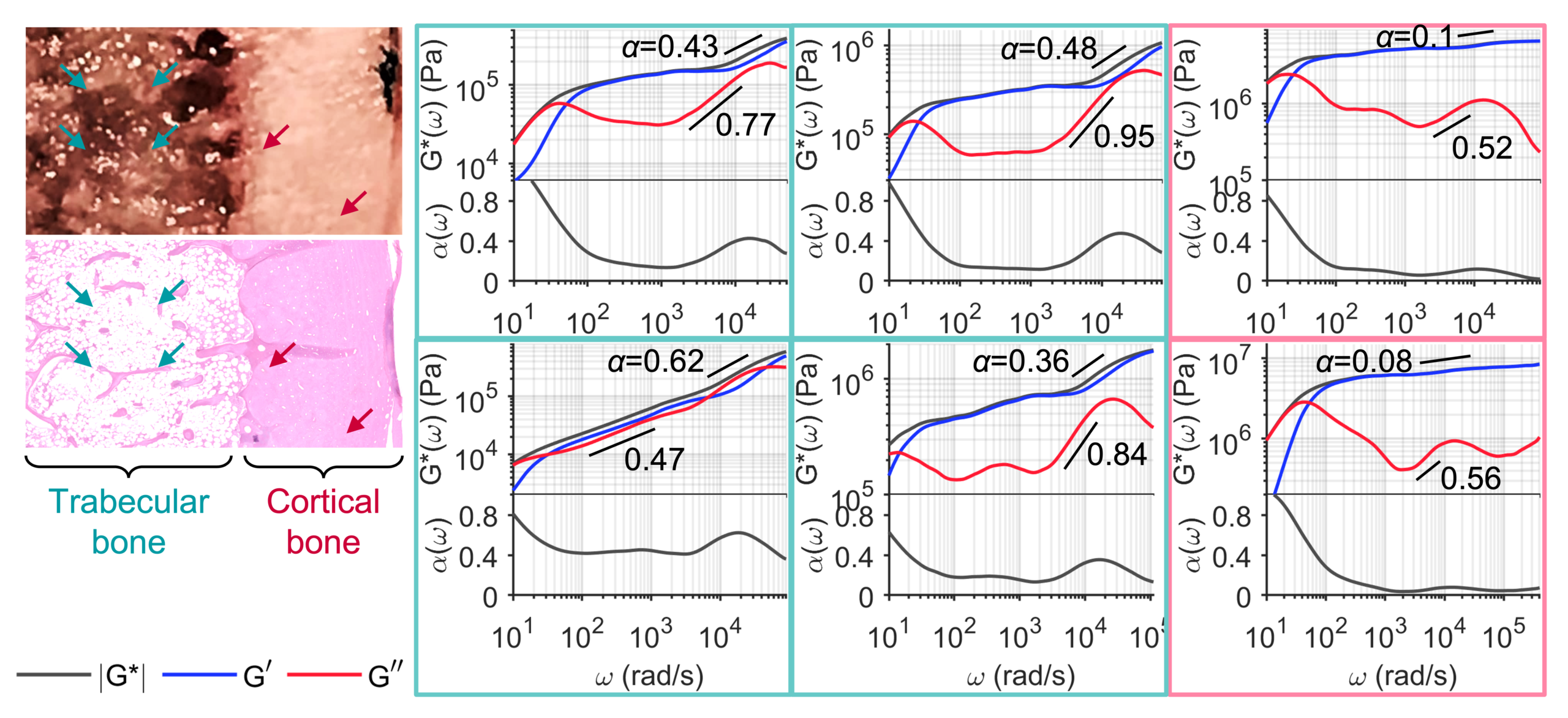
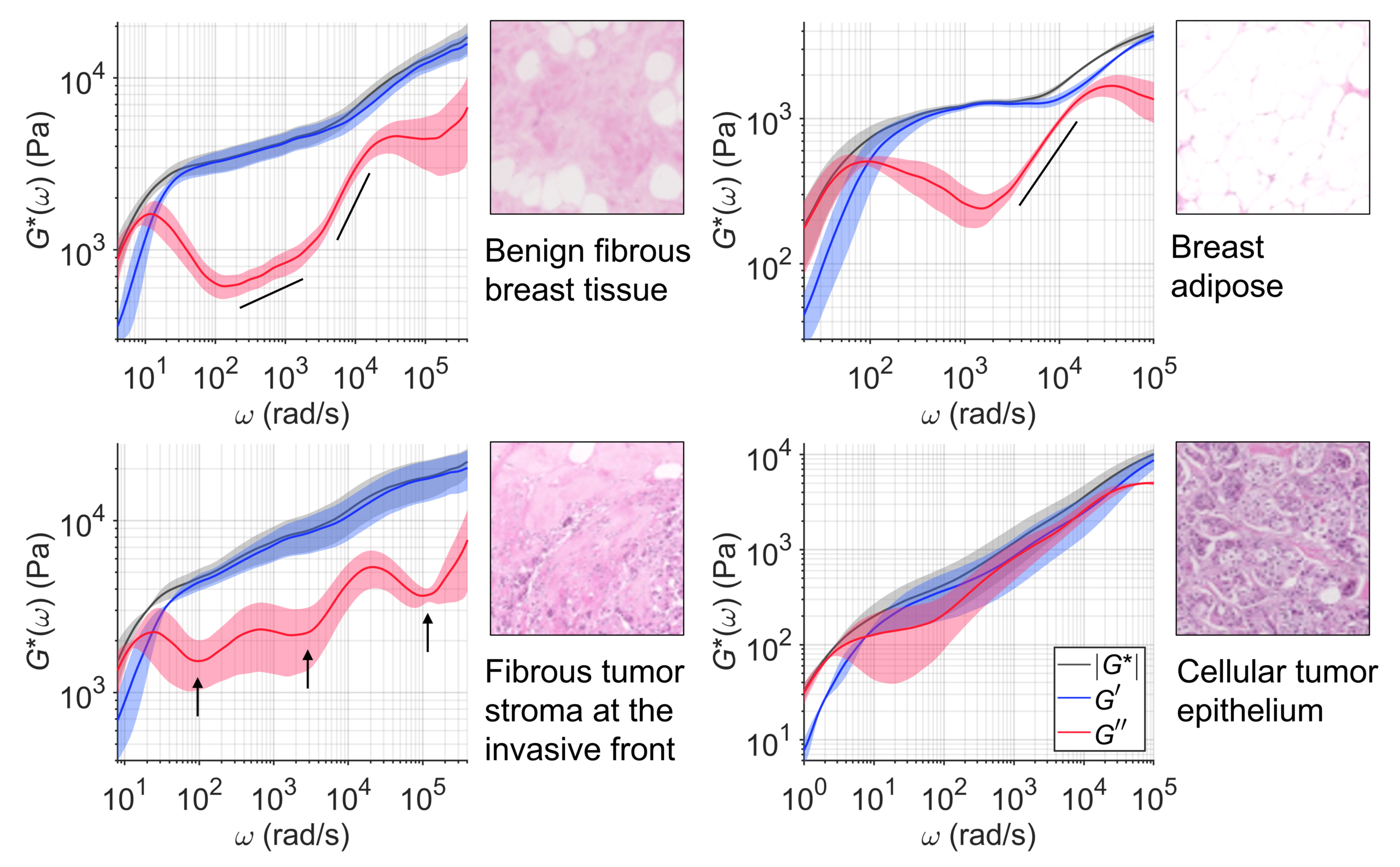
Biological tissues scale a broad range of mechanical properties, from pliant fatty tissue to hard bone. Abnormal changes in tissue mechanics are a powerful driver of various diseases including cancer, fibro-proliferative disorders, hematological disorders, and orthopedic conditions. Thus, mechanical signatures of tissue can enhance our understanding of the disease state and improve our ability to diagnose and treat diseases. However, current clinical pathology procedures do not comprehensively discern mechanical abnormalities of clinical tissue. Our lab develops a new tool in the form of a simple upright microscope, called SHEAR, to enable high-resolution mapping of tissue micromechanics in gross unprocessed tissue specimens. Of note, SHEAR has established the links between microscale heterogeneities of viscoelasticity and histopathological subtype, tumor grade, receptor expression, and lymph node status in breast carcinoma. Our other application areas include tissue engineering and regenerative medicine of knee joint tissues andatherosclerotic plaque biomechanics.

Micromechanical mapping of the breast tumor microenvironment

Of note, SHEAR measurements have established the links between microscale heterogeneities of viscoelasticity and histopathological subtype, tumor grade, receptor expression, and lymph node status in breast carcinoma. By providing detailed information about the mechanical properties of lesions, SHEAR has the potential to greatly enhance our understanding of the disease mechano-pathology and improve our ability to diagnose and treat diseases.

Wideband (WB)-SHEAR

Mechanical properties of biological issues are not merely a single fixed value of modulus. Both the elastic and viscous moduli are dynamically modulated over the frequency at which the tissue is being probed, yielding fingerprint-like frequency-dependent viscoelastic spectra that span a wide frequency band. Living cells, in fact, sense and respond to elastic and viscous mechanical cues at distinct frequencies ranging from sub-Hz to sub-MHz, depending on their activity and how fast they interact with the microenvironment. To capture these intricate multiscale behaviors, we develop a wideband variant of SHEAR (WB-SHEAR) that utilizes an ultra-high-speed camera to probe shear elastic and viscous moduli up to the sub-MHz regime.  These wideband ‘viscoelastic fingerprints’ uniquely characterize tissue micromechanics in far more comprehensive ways than a typical measurement of stiffness at a single frequency.


Micromechanical mapping of the frequency-dependent
wideband viscoelastic spectra in breast tumor. Right: Frequency-dependent maps
of G′(ω) (top) and G″(ω) (bottom). Scale bar: 50
μm. Left: Frequency-dependent |G*(ω)| (black), G′(ω)
(blue), and G″(ω) (red) spectra at the locations indicated by +
(top) and ○ (bottom) on the maps.